Radiology AI Foundation Models: Promising But Not Ready
A new review of 67 studies found that radiology-specific vision foundation models transfer well between tasks - but rarely get tested across hospitals and scanners. That missing test is exactly what stands between promising research and clinical use.
Automation needs a narrow first win
The best first AI workflow is usually a repeated task with a clear input, clear output, and a human approval step.
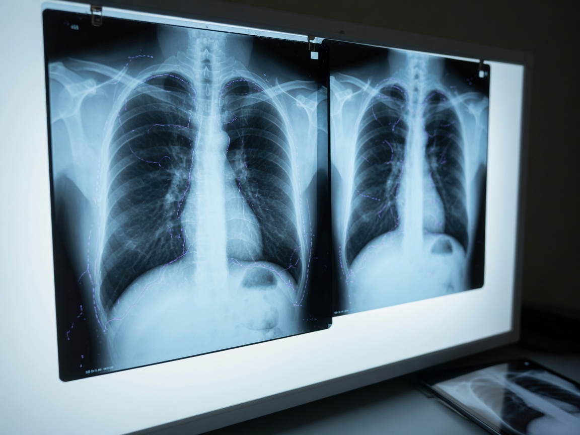
A vision foundation model for radiology is an AI system pretrained on huge volumes of medical scans - brain MRIs, chest X-rays, CT images - so it can later be adapted to many specific tasks like spotting or outlining abnormalities. A new scoping review of 67 studies, published between January 2017 and March 2026, took stock of these models, and its one-sentence conclusion is worth sitting with: they show promising transferability, but clinical translation remains constrained.
What The Review Actually Found
The researchers behind this review mapped radiology-specific vision foundation models across three pillars: how big and varied the training data is, how the architectures and pretraining scale, and how well the models transfer to new tasks.
The datasets leaned heavily toward brain MRI, thoracoabdominal CT, and chest X-ray, and their sizes ranged from fewer than 100,000 samples to multi-million-image cohorts. On the technical side, transformer-based architectures dominated, almost always paired with self-supervised pretraining - techniques like masked image modeling (where the model learns by filling in hidden patches of an image), contrastive learning (learning by telling similar images apart from different ones), and multi-stage approaches that combine methods.
When it came to testing, most studies measured segmentation (tracing structures pixel by pixel) and classification (labeling a scan as one thing or another). That's where the pattern gets interesting.
Worth a look
Phugialy Picks

LLM Systems Engineering: Training and Building Large Language Models – Engineering AI Models Through Fine-Tuning, Continued Pretraining, and From-Scratch Development (AI Engineering)

Anker USB-C to USB-C 60W Fast Charging Cable (2-Pack)
We'd buy this if: You need a reliable spare charging cable that won't fray in a month.
We'd skip this if: You already have enough cables lying around.
Stanley Quencher H2.0 Tumbler 30oz
We'd buy this if: You want a large, well-reviewed insulated tumbler and don't mind it having nothing to do with AI.
We'd skip this if: You'd rather every Pick on this site stay strictly on-topic.
Some Phugialy Picks use affiliate links. If you buy through one, Phugialy may earn a commission. It doesn't change what we recommend. Full disclosure →
The Gap Between Lab Scores And Clinical Reality
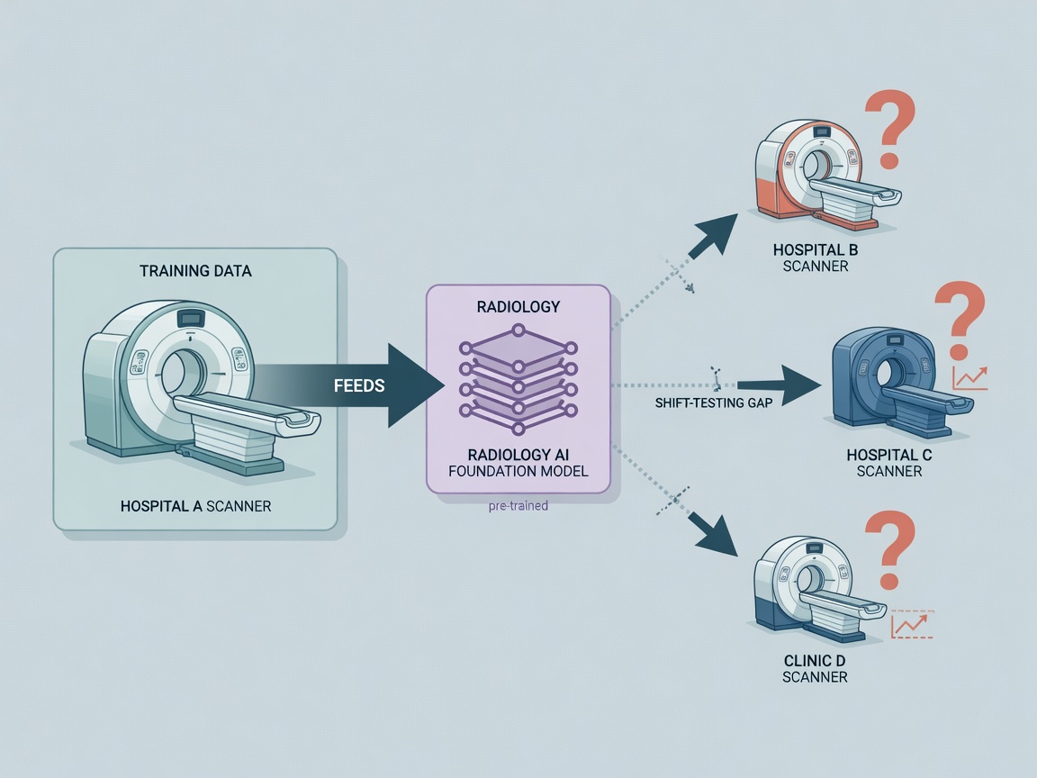
Here's the part most coverage glosses over: a model can ace segmentation benchmarks in one hospital's data and still fail quietly somewhere else. The review found that validation under real-world shifts - cross-center differences, cross-scanner differences, changes in anatomy or imaging modality - was inconsistently reported. In other words, we often don't know whether these models generalize beyond the exact conditions they were trained in.
It's easy to conflate "good benchmark performance" with "ready for the clinic," but they're not the same thing. The review names four specific constraints: limited data representativeness (the training scans don't reflect the full diversity of real patients and machines), heterogeneous benchmarks (every study measures differently), incomplete reporting (you can't audit what wasn't written down), and insufficient deployment-oriented evaluation (almost nobody tests these models the way they'd actually run in a hospital workflow).
What This Actually Points To
The real story here isn't that radiology foundation models are overhyped - it's that the field is measuring them with tools built for a different question. "Can this model segment tumors well on this dataset?" is a solvable research question. "Will this model behave safely on a scanner from a different vendor at a hospital it's never seen?" is the clinical question, and per this review, it's rarely being asked.
My read as an interpretation: the promising transferability results are genuinely encouraging - self-supervised pretraining on large radiological cohorts does seem to produce models worth adapting. But until shift-testing becomes standard reporting rather than an optional extra, every benchmark number should come with an invisible asterisk. The bottleneck isn't architecture anymore; it's evidence.
Got a question about how this applies to you? →
Keep reading
Follow the thread
Mamba-Based Deep Clustering for Time Series: Real or Hype?
A new Mamba-based framework claims linear-complexity deep clustering for time series, winning 29 of 60 metric evaluations across 15 benchmarks. The architecture is genuinely well-aimed at the unlabeled-data problem — but benchmark wins aren't production evidence.
Read this noteSame lane, different angle
Multimodal Federated Learning Just Got Better at Missing Data
Hospitals could train shared diagnostic models without patient data ever leaving their walls — even when one site's imaging setup can't capture everything. A new framework called ProMoE-FL synthesizes the missing pieces on the fly.
AnchorPrune: Training-Free Token Pruning That Almost Holds Up
A training-free framework called AnchorPrune keeps 97.6% of full-token performance in a vision-language model using only 160 of 2,880 visual tokens - no retraining required. The interesting part isn't the compression ratio; it's what gets protected first.